The role of the periaqueductal gray (PAG) has been described in many studies regarding pain processing. This structure, located in the midbrain around the cerebral aqueduct, is mainly known as a key region in descending pain modulation. However, the PAG is related to a wide spectrum of functions, such as pain, analgesia, fear, anxiety, and cardiovascular functions. Furthermore, the PAG has been a target in chronic pain research, whereas specific brain abnormalities in the PAG were found [Mainero et al. 2011; Desouza et al. 2013]. PAG functional connectivity has also been shown adaptive to treatment in chronic pain patients [Erpelding et al. 2014].
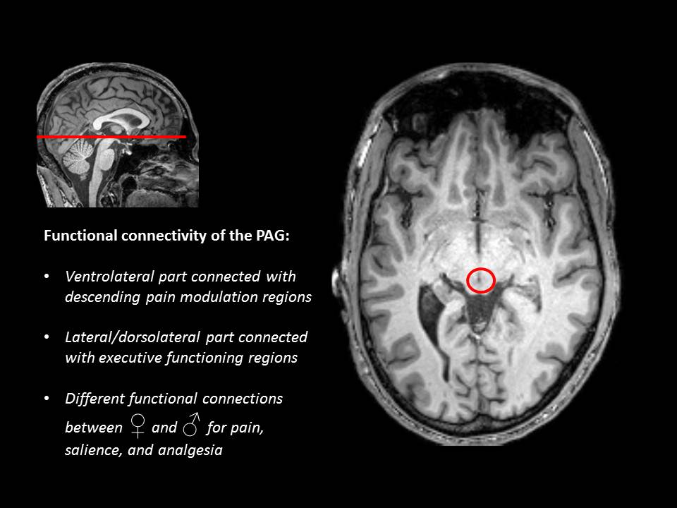
In addition to the multiple functions of the PAG, a new study of Coulombe et al. (2016) showed that functional connectivity of sub-regions of the PAG have different roles in pain and pain coping, yet showed also differences between men and women. In a resting-state fMRI experiment, Coulombe and colleagues found that the ventrolateral part of the PAG was connected to brain regions associated with descending pain modulation, whereas the lateral and dorsolateral PAG were connected to brain regions implicated in executive functioning. In addition, they found different functional connections of the PAG to brain regions involved in pain, salience, and analgesia in men and women.
To conclude, specific sub-regions of the PAG are involved in descending pain modulation and executive functioning, while there is also a gender difference in functional connectivity from the PAG to other brain regions. These findings may play a role in expanding our knowledge of the underlying pathology of chronic pain and corresponding coping mechanisms.
Jeroen Kregel
2016 Pain in Motion
References and further reading:
Mainero C, Boshyan J, Hadjikhani N. Altered functional magnetic resonance imaging resting-state connectivity in periaqueductal gray networks in migraine. Ann Neurol. 2011 Nov;70(5):838-45. doi: 10.1002/ana.22537.
http://www.ncbi.nlm.nih.gov/pmc/articles/PMC3243965/
Desouza DD, Moayedi M, Chen DQ, Davis KD, Hodaie M. Sensorimotor and Pain Modulation Brain Abnormalities in Trigeminal Neuralgia: A Paroxysmal, Sensory-Triggered Neuropathic Pain. PLoS One. 2013 Jun 18;8(6):e66340. Print 2013.
http://www.ncbi.nlm.nih.gov/pmc/articles/PMC3688879/
Erpelding N, Simons L, Lebel A, Serrano P, Pielech M, Prabhu S, Becerra L, Borsook D. Rapid treatment-induced brain changes in pediatric CRPS. Brain Struct Funct. 2016 Mar;221(2):1095-111. doi: 10.1007/s00429-014-0957-8.
http://www.ncbi.nlm.nih.gov/pubmed/25515312
Coulombe MA, Erpelding N, Kucyi A, Davis KD. Intrinsic functional connectivity of periaqueductal gray subregions in humans. Hum Brain Mapp. 2016 Apr;37(4):1514-30. doi: 10.1002/hbm.23117.